Your Highlands Ranch Eye Doctor
Located off of C470 and Quebec in the King Soopers Parking lot.
About Highlands Ranch HD Eye Care
At Highlands Ranch HD Eye Care, we are committed to providing a high level of personalized care to improve your vision and protect your eye health. Our positive attitude towards life and our patients is the foundation of what makes our practice the best choice for your family. We believe that healthy eyesight is vital to a high quality of life, and to “enhance and preserve the gift of sight” is the essence of our practice philosophy. We look forward to helping you and your family find the best pair of glasses, sunglasses, contact lenses, Myopia Management, Orthokeratology (Ortho eyes), or Lasik to meet your visual needs.
In everything we do, we provide an excellent experience for our patients with a truly caring staff and personal HD products that help everyone see better, look better and play better.


Our Eye Care Services

Comprehensive Eye Exams
From updating your eyeglasses prescription to detection and treatment of eye diseases, comprehensive eye exams are important for continued visual health.

Contact Lens Exams
During a contact lens exam, your eye doctor will check if you are a good candidate for contacts and find the best type of contacts for your needs..

Dry Eyes
Do you have dry, itchy, gritty-feeling eyes? Dry eye syndrome is a very common condition. We offer dry eye treatments at our eye care clinic.
Clear vision is an essential part of a child’s healthy development and learning. Regular pediatric eye exams can help detect and treat common issues.
Children with myopia are at higher risk for potentially sight-threatening vision problems. Save your child’s vision with myopia management.
Eye diseases such as glaucoma and macular degeneration can cause severe loss of vision if not diagnosed and treated early. Our eye care team can help.
LASIK and refractive surgery are great ways to say goodbye to contacts and glasses for good. Find out if you’re a good candidate.
If cataracts go untreated, they can cause total blindness in the affected eye. We can help with co-management of your cataract removal surgery.
Astigmatism can cause vision problems such as blurred or double vision at all distances. Find out what you can do to correct astigmatism and see your best.
Macular degeneration can cause severe central vision loss if not detected and treated early on. We can help preserve your vision.
Over 50% of people living with glaucoma don’t know they have the disease as it shows no symptoms. Early detection and treatment can prevent blindness.
The ups and downs of diabetes can be difficult to navigate. How can diabetes affect your eyes? How can you and your eye doctor keep them healthy?
Presbyopia is a normal part of aging. If you’re past 40 and notice you can’t read up close, our eye doctors may be able to help.
InfantSEE is a program that provides free comprehensive eye exams to infants 6-12 months old, regardless of a family’s financial status.
While vision can change drastically in the senior years, these changes shouldn't have to impair one's quality of life. The earlier these problems are detected and treated, the more likely proper eye care can help you retain good vision.
Orthokeratology is a non-invasive vision correction alternative to LASIK. Put in the specialty lenses while you sleep, and experience better vision in the morning.
Eyeglasses & Frames

Designer Frames
Our extensive optical section offers a wide variety of eyeglass frames in every style, material & design. Come visit us today to see for yourself!
Our expert optical team can find just the right pair of glasses for you to be confident and look your best.
Lens coatings improve visual comfort, make it easier to clean your glasses and ensure your lenses last longer. Coatings include anti-scratch, anti-reflective, photochromatic and UV / blue light filters.
Contact Lenses

Contact Lens Fitting
We offer a wide range of contact lens options from dailies, monthly to multifocal contact lenses for crystal clear vision and superior comfort.
During a contact lens exam, your eye doctor will check if you are a good candidate for contacts and find the best type of contacts for your needs.
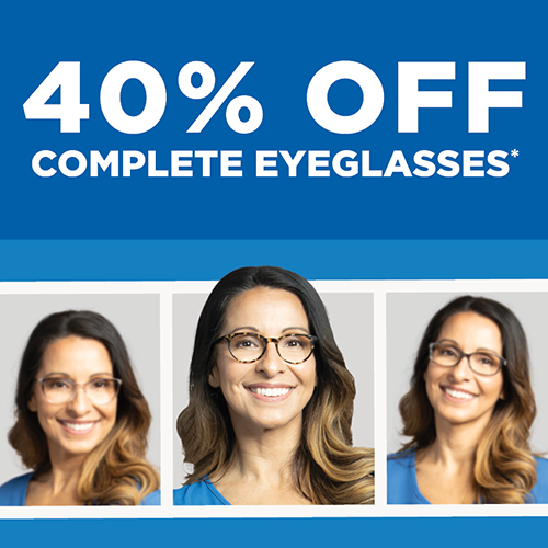
40% Off Complete Eyeglasses.
*Requires purchase of a complete prescription pair, including frame and lenses. Does not include sunglass frames, Barton Perreira, Cartier, Cazal, Chanel, Cutler and Gross, Dior, Dita Lancier, Fendi, Gucci, ic!Berlin, l.a. Eyeworks, Maui Jim, Mykita, Nifties, Oakley, Oliver Peoples, Persol, Ray-Ban, Robert Marc, Salt, Salvatore Ferragamo, Skaga, Silhouette, Tom Ford, WOOW, accessories, contact lenses, or medical procedures. Cannot be combined with any other discounts, promotions, or insurance plans. Not valid on previous orders. Other restrictions may apply. See practice for full details. Offer valid 04/08/2024-06/16/2024. 24AEG-729313